
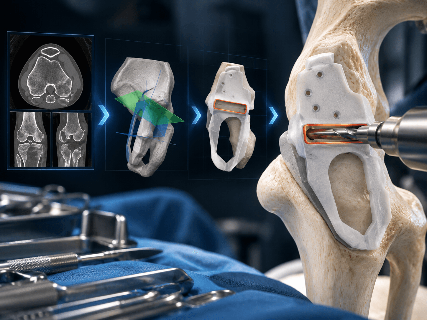
Engineered for the OR.
Rigorous translation from DICOM multislice CT datasets to 3D virtual surgical plans, patient specific cutting guides, and operative outcomes.
End to End Engineering Workflows.
Documented surgical cases demonstrating initial radiologic scan, multiplane virtual osteotomy, patient matched instrument design, and physical verification.
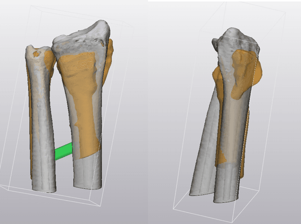
Distal Radius Malunion Corrective Osteotomy
Clinical Presentation: Symptomatic extra articular distal radius deformity following fracture malunion.
Cost effective patient specific osteotomy guide incorporating pre drilled screw trajectories and planned volar plate osteoplasty, validating correction against contralateral mirrored anatomy with standard VA LCP reduction.
Thin slice CT acquisition; volumetric segmentation of malunited radius, ulna, and contralateral normal forearm for mirror template generation.
3D multiplanar realignment targeting physiological values: Radial Inclination (14.75° → 21.9°), Volar Tilt (28.47° → 15.61°), and Radial Height (7.0mm → 11.0mm).
Engineered patient matched contact guide with saw blade slot, standard VA LCP screw trajectories, and secondary plate osteoplasty resection slot.
Physical 3D printing of bone bed, custom stepped spacers, and custom guide; successful reduction achieved using commercially available anatomical locking plates.
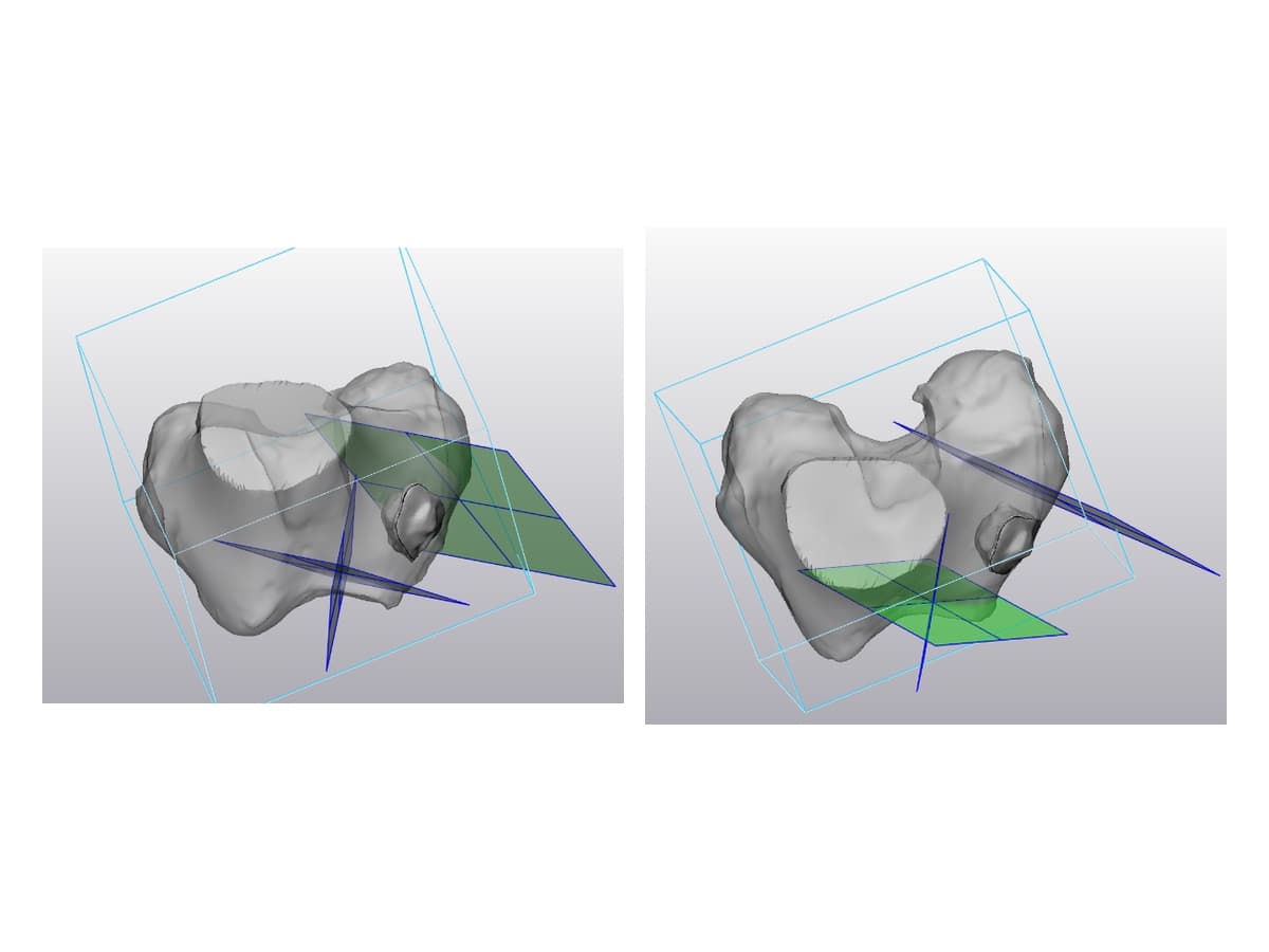
Distal Femoral Resection & Saw Slot Capture
Clinical Presentation: Distal femoral deformity and joint alignment failure requiring precision single cut wedge resection.
Patient matched distal femoral cutting block designed with rigid anatomical wraparound fit, dual plane saw containment, and cortical reference pins to eliminate blade chatter.
High resolution DICOM scanning with submillimeter axial slicing and dynamic cortical density thresholding.
Virtual anatomical to mechanical axis realignment with calibrated cut planes defined in axial, medial, and anteromedial perspectives.
Monolithic patient specific cutting block incorporating rigid registration flanges, captive saw slots, and anterior anchor pin channels.
Full scale anatomical bone replicate 3D printed to verify zero intraoperative slip and confirm tactile guide seating prior to surgical clearance.
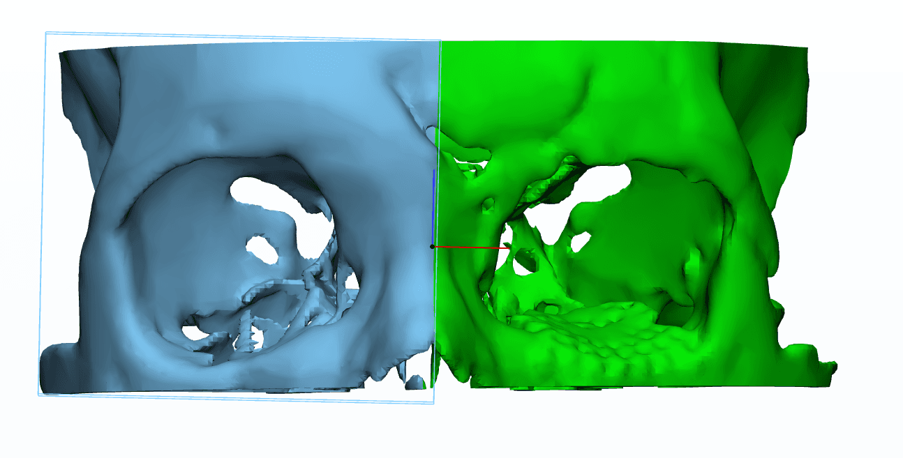
Supraorbital Ridge & Orbital Floor Trauma Reconstruction
Clinical Presentation: Post traumatic craniofacial deformity with persistent enophthalmos following prior titanium mesh placement.
Computer aided mirror reconstruction and 3D surface deviation mapping evaluating posteroinferior orbital floor deficiencies to guide custom multiple size anatomical augments.
0.625mm helical CT scan parsing maxilla, zygoma, and bilateral orbits; segmentation via Mimics 21.
Unaffected contralateral orbit mirrored; global registration reduced initial 12.2mm deviation down to 5.64mm on the supraorbital rim.
Color coded surface deviation mapping identified critical absence of posteroinferior bony ledge underlying the previous titanium mesh.
Virtual augmentation design generating 3 to 4 graduated patient matched implants for optimal intraoperative volume restoration.
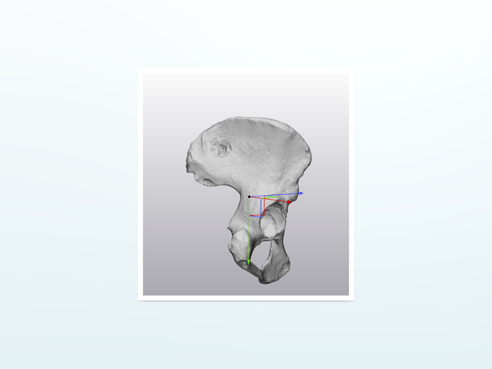
Internal Hemipelvectomy & Sacral Ala Resection
Clinical Presentation: Primary bone sarcoma and pelvic tumor demanding clear surgical resection margins while shielding the sacral nerve roots.
Multiplane custom pelvic osteotomy guides achieving sub 2mm margin fidelity; postoperative CT surface comparison validated 88% of resection area within ±2.5mm.
Multiparametric CT volume extraction identifying intrapelvic lesion boundaries and bone soft tissue interfaces.
Supraacetabular and posterior iliac osteotomy planes engineered to safeguard sacral nerve roots and critical sciatic neurovascular bundles.
Primary seating guide matched to posterior ilium anatomy with secondary drill templates to ensure exact mechanical screw trajectories.
Postoperative CT global surface comparison confirmed 78% of cuts within ±2mm with clear oncological resection margins.
Have a patient scan ready for 3D guide feasibility?
Upload anonymous DICOM parameters for virtual osteotomy planning and custom guide verification.